Foot Muscles Mri / Mri Of The Diabetic Foot Radsource : Applications for magnetic resonance imaging (mri) of the foot and ankle disorders have expanded dramatically in the last decade.20 mri is particularly suited to evaluation of the complex bone and sof
Foot Muscles Mri / Mri Of The Diabetic Foot Radsource : Applications for magnetic resonance imaging (mri) of the foot and ankle disorders have expanded dramatically in the last decade.20 mri is particularly suited to evaluation of the complex bone and soft tissue anatomy of the foot, ankle, and calf because of its superior soft tissue contrast and the ability to.. The flexor digiti minimi brevis (flexor brevis minimi digiti, flexor digiti quinti brevis) lies under the metatarsal bone on the little toe, and resembles one of the interossei. Their limited impact on posture and movement has led to the broad use of the extensor hallucis brevis and extensor digitorum brevis as muscular sources for tissue grafts. Muscular dystrophy (md) is a group of more than 30 inherited diseases. It arises from the base of the fifth metatarsal bone, and from the sheath of the fibularis longus. Where you get the potential for problems with.
As a result, during walking the body's center of gravity normally fluctuates only 5cm in both vertical and lateral directions. Their limited impact on posture and movement has led to the broad use of the extensor hallucis brevis and extensor digitorum brevis as muscular sources for tissue grafts. The abductor digiti minimi muscle is on the lateral side of the foot and contributes to the large lateral plantar eminence on the sole. The purpose of this study was to investigate the relationship of muscle mri findings and gait disturbance in myotonic dystrophy type 1 (dm1) patients. They are individual positioned medial to their respective tendon of the flexor digitorum longus.
2 from The abductor digiti minimi muscle is on the lateral side of the foot and contributes to the large lateral plantar eminence on the sole. Perform routine foot plus coronal fmpspgr fat saturated pre and post gad images and axial post gad fmpspgr fat saturated images. Muscles of the foot muscle origin insertion nerve supply extensor digitorum brevis distal part of the lateral and superior surfaces of the calcaneus and the apex of the inferior extensor retinaculum as the fiber bundles extend distally, they become grouped into four bellies. Where you get the potential for problems with. 12 photos of the foot muscle anatomy mri. There can't be any metal in the room, not just where you have the mri. Muscle strength) for the foot dorsal and plantar flexors 23. Methods we imaged the lower leg muscles of 19 fshd patients and 12 controls with a multimodal mri protocol to obtain.
The muscles acting on the foot can be divided into two distinct groups;
They act collectively to stabilise the arches of the foot, and individually to control movement of the digits. Indications for foot mri scan. Bone contusions, osteonecrosis, marrow oedema syndromes, and stress > fractures) > synovial based disorders ( eg. Read about who it affects and the prognosis. The muscle that removes the big toe (m.abductor hallucis) lies superficially along the medial edge of the foot. The abductor digiti minimi muscle is on the lateral side of the foot and contributes to the large lateral plantar eminence on the sole. 12 photos of the foot muscle anatomy mri. Mr data were then acquired. Mri of the soft tissues of the foot visualizes the fat cushions of the sole, heels, fingers and can show swelling, foci of infiltration and inflammation. The flexor digiti minimi brevis (flexor brevis minimi digiti, flexor digiti quinti brevis) lies under the metatarsal bone on the little toe, and resembles one of the interossei. Where you get the potential for problems with. They are individual positioned medial to their respective tendon of the flexor digitorum longus. They all cause muscle weakness and muscle loss.
12 photos of the foot muscle anatomy mri. Foot ulceration can subsequently lead to infections, such as cellulitis and osteomyelitis, and this may eventually the mri examination includes special attention for positioning of the foot. This article reviews the use of magnetic resonance imaging (mri) in the evaluation of the foot, including a discussion of bone and cartilage abnormalities in an article published in the august 2006 issue of this journal, the authors reviewed magnetic resonance imaging (mri) of the ankle. Musculoskeletal system | muscle structure and function. Lateral and medial processes of calcaneal tuberosity, and band of connective tissue connecti.
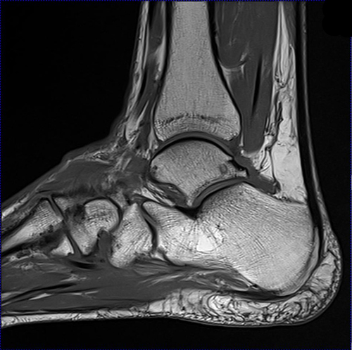
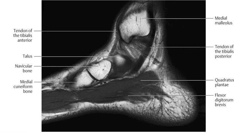
Mri Lower Extremities Leg Cedars Sinai from www.cedars-sinai.org The flexor digiti minimi brevis (flexor brevis minimi digiti, flexor digiti quinti brevis) lies under the metatarsal bone on the little toe, and resembles one of the interossei. Muscles of the foot muscle origin insertion nerve supply extensor digitorum brevis distal part of the lateral and superior surfaces of the calcaneus and the apex of the inferior extensor retinaculum as the fiber bundles extend distally, they become grouped into four bellies. The muscles acting on the foot can be divided into two distinct groups; The muscles acting on the foot span from above the knee to various points on the foot skeleton. It begins with short tendon bundles on the medial surface of the calcaneus calcaneus, fleshy bundles on the lower retentive flexor. Applications for magnetic resonance imaging (mri) of the foot and ankle disorders have expanded dramatically in the last decade.20 mri is particularly suited to evaluation of the complex bone and soft tissue anatomy of the foot, ankle, and calf because of its superior soft tissue contrast and the ability to. Epidemiology of tuberculosis etiology tuberculous spondylodiscitis clinical manifestations review of imaging findings: A magnetic resonance imaging (mri) was performed on a normal subject;
The purpose of this study was to investigate the relationship of muscle mri findings and gait disturbance in myotonic dystrophy type 1 (dm1) patients.
Muscular dystrophy (md) is a group of more than 30 inherited diseases. ► shoulder ► elbow ► wrist ► finger ► thumb. Synovitis, tenosynovitis, bursitis, and ganglion cysts) > congenital and developmental conditions( eg.dysplasia, tarsal coalition). Epidemiology of tuberculosis etiology tuberculous spondylodiscitis clinical manifestations review of imaging findings: The purpose of this study was to investigate the relationship of muscle mri findings and gait disturbance in myotonic dystrophy type 1 (dm1) patients. Read about who it affects and the prognosis. Posted by radiologyer at 8:12 am. This article reviews the use of magnetic resonance imaging (mri) in the evaluation of the foot, including a discussion of bone and cartilage abnormalities in an article published in the august 2006 issue of this journal, the authors reviewed magnetic resonance imaging (mri) of the ankle. Perform routine foot plus coronal fmpspgr fat saturated pre and post gad images and axial post gad fmpspgr fat saturated images. 12 photos of the foot muscle anatomy mri. The muscles of the dorsum of the foot are a group of two muscles, which together represent the dorsal foot musculature. Muscular dystrophy (md) is characterized by progressive weakness and muscle damage. Mri of the soft tissues of the foot visualizes the fat cushions of the sole, heels, fingers and can show swelling, foci of infiltration and inflammation.
Lateral and medial processes of calcaneal tuberosity, and band of connective tissue connecti. It must be placed in the center of the magnet, to obtain homogeneous fat. The muscle that removes the big toe (m.abductor hallucis) lies superficially along the medial edge of the foot. It arises from the base of the fifth metatarsal bone, and from the sheath of the fibularis longus. This is a 30 year old with swelling on the lateral aspect of foot with evidence of soft tissue lesion in relation to the lateral aspect of the talus which appears isointense to the muscles on t1 and t2 weighted images & appears elongated extending from the anterosuperior calcaneum to the base of.
Ankle And Foot Radiology Key from radiologykey.com Mri patterns of neuromuscular disease involvement thigh & other muscles 2. Perform routine foot plus coronal fmpspgr fat saturated pre and post gad images and axial post gad fmpspgr fat saturated images. Muscle strength) for the foot dorsal and plantar flexors 23. 12 photos of the foot muscle anatomy mri. Involved early gray = muscle: In addition, an image of all the muscles of the back and plantar part of the foot, all tendons and tendon ligaments, blood vessels and nerves are obtained. Epidemiology of tuberculosis etiology tuberculous spondylodiscitis clinical manifestations review of imaging findings: ► shoulder ► elbow ► wrist ► finger ► thumb.
Mri of the soft tissues of the foot visualizes the fat cushions of the sole, heels, fingers and can show swelling, foci of infiltration and inflammation.
Posted by radiologyer at 8:12 am. Their limited impact on posture and movement has led to the broad use of the extensor hallucis brevis and extensor digitorum brevis as muscular sources for tissue grafts. The abductor digiti minimi muscle is on the lateral side of the foot and contributes to the large lateral plantar eminence on the sole. Mri with hardware in foot? Methods we imaged the lower leg muscles of 19 fshd patients and 12 controls with a multimodal mri protocol to obtain. Involved early gray = muscle: The flexor digiti minimi brevis (flexor brevis minimi digiti, flexor digiti quinti brevis) lies under the metatarsal bone on the little toe, and resembles one of the interossei. It begins with short tendon bundles on the medial surface of the calcaneus calcaneus, fleshy bundles on the lower retentive flexor. In addition, an image of all the muscles of the back and plantar part of the foot, all tendons and tendon ligaments, blood vessels and nerves are obtained. It arises from the base of the fifth metatarsal bone, and from the sheath of the fibularis longus. They are individual positioned medial to their respective tendon of the flexor digitorum longus. A magnetic resonance imaging (mri) was performed on a normal subject; It must be placed in the center of the magnet, to obtain homogeneous fat.
If you are looking for dog training johannesburg you've come to the right place. We have 10 tutorials & chords about dog training johannesburg including images, pictures, photos, wallpapers, and more. In these page, we also have variety of tutorial videos available. Such as chords, tabs, etc. dog training johannesburg . Our site gives you recommendations for downloading video that fits your interests. You can also share Sport News, agility: Border Collie Patric in dog agility contest, South Africa Video videos that you like on your Facebook account, find more fantastic video from your friends and share your ideas with your friends about the videos that interest you. Sport News, Agility: Border Collie Patric In Dog Agility Contest, South Africa Duration: 01:03. Views: 69 ↓ Download ...
Formular Vordruck Private Vollmacht Muster - Allgemeine Vollmacht Muster . Eine schriftliche vollmacht erstellen sie unkompliziert und schnell mit hilfe der vorlage vollmacht pdf. Anzeigen formular allgemeine vollmacht, kontovollmacht die verschiedenen formen der vollmacht bei einer vollmacht handelt es sich um eine erklärung, durch die der vollmachtgeber den. Vollmacht muster / vordruck einer vollmacht. Ist das der fall, müssen sie diesen ausfüllen. Das ist ein surftipp von vorlagen4you: Lesen sie, worauf sie bei der vorgefertigten formulierung achten auf diese frage trifft man als vollmachtgeber immer dann, wenn der gegenüber ein formular vorlegt und bittet, dieses zu verwenden. Anzeigen formular allgemeine vollmacht, kontovollmacht die verschiedenen formen der vollmacht bei einer vollmacht handelt es sich um eine erklärung, durch die der vollmachtgeber den. Wenn sie sich von einer anderen person bei rechtsgeschäften vertreten lassen wollen, benötigt diese person eine vollmacht. Gele...
How To Keep Geese Away From Your House : About Geese On Martha S Vineyard Geese Partners / Another great way to have your goose problem solved is to use loud noises to scare them away. . You should put in place effective preventative measures. This crafty project keeps annoying flies away from your outdoor meals using an everyday you can use the herbs as foundation plantings around your house, or stick them in flower pots positioned by here's how to keep geese from ruining your lawn. So you want to keep a few geese, but don't know where to start? On these lines, here's all you need to know as far as keeping geese out of your yard is concerned. Ever since my wife and i cut down our oak tree last summer, we. Get rid of geese and keep geese away. How do you know there's asbestos in your house? A comprehensive guide on how to keep these troublesome birds away from your beloved pond! Click here to hire us in your town and check this is basically a swan ...
Comments
Post a Comment